Cardiovascular issues are rapidly becoming a key danger in coronavirus disease 2019 (COVID-19) in addition to breathing illness. The mechanisms underlying the out of proportion effect of extreme intense respiratory syndrome coronavirus 2 (SARS-CoV-2) infection on patients with cardiovascular comorbidities, however, stay incompletely comprehended.
1
- Zhou F
- Yu T
- Du R
- et al.
Medical course and risk aspects for mortality of adult inpatients with COVID-19 in Wuhan, China: a retrospective friend research study.
,
2
- Horton R
Offline: COVID-19– confusion and candour.
SARS-CoV-2 contaminates the host using the angiotensin transforming enzyme 2 (ACE2) receptor, which is revealed in numerous organs, consisting of the lung, heart, kidney, and intestine. ACE2 receptors are likewise expressed by endothelial cells.
3
- Ferrario CM
- Jessup J
- Chappell MC
- et al.
Impact of angiotensin-converting enzyme inhibition and angiotensin II receptor blockers on cardiac angiotensin-converting enzyme 2.
Whether vascular derangements in COVID-19 are due to endothelial cell involvement by the infection is currently unidentified. Intriguingly, SARS-CoV-2 can straight contaminate engineered human capillary organoids in vitro.
4
- Monteil V KH
- Prado P
- Hagelkrüys A
- et al.
Inhibition of SARS-CoV-2 infections in crafted human tissues utilizing clinical-grade soluble human ACE2.
Here we show endothelial cell participation across vascular beds of various organs in a series of clients with COVID-19(additional case details are offered in the appendix).
Patient 1 was a male renal transplant recipient, aged 71 years, with coronary artery illness and arterial high blood pressure. The patient’s condition degraded following COVID-19 medical diagnosis, and he needed mechanical ventilation. Multisystem organ failure happened, and the patient passed away on day 8.
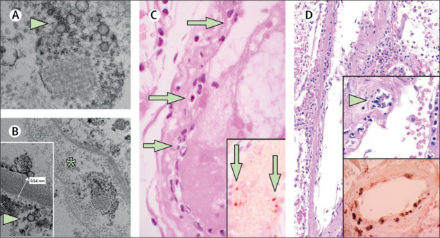
Post-mortem analysis of the transplanted kidney by electron microscopy exposed viral inclusion structures in endothelial cells (figure A, B). In histological analyses, we found an accumulation of inflammatory cells connected with endothelium, along with apoptotic bodies, in the heart, the small bowel (figure C) and lung (figure D). An accumulation of mononuclear cells was discovered in the lung, and the majority of small lung vessels appeared congested.

Figure Pathology of endothelial cell dysfunction in COVID-19
Show complete caption
( A, B) Electron microscopy of kidney tissue shows viral addition bodies in a peritubular space and viral particles in endothelial cells of the glomerular capillary loops. Aggregates of viral particles (arrow) appear with thick circular surface and lucid centre. The asterisk in panel B marks peritubular space constant with capillary consisting of viral particles. The inset in panel B shows the glomerular basement membrane with endothelial cell and a viral particle (arrow; about 150 nm in diameter). (C) Small bowel resection specimen of patient 3, stained with haematoxylin and eosin. Arrows indicate dominant mononuclear cell infiltrates within the intima along the lumen of lots of vessels. The inset of panel C shows an immunohistochemical staining of caspase 3 in little bowel specimens from serial section of tissue explained in panel D. Staining patterns were consistent with apoptosis of endothelial cells and mononuclear cells observed in the haematoxylin-eosin-stained sections, indicating that apoptosis is induced in a substantial proportion of these cells. (D) Post-mortem lung specimen stained with haematoxylin and eosin showed thickened lung septa, consisting of a large arterial vessel with mononuclear and neutrophilic seepage (arrow in upper inset). The lower inset shows an immunohistochemical staining of caspase 3 on the same lung specimen; these staining patterns were consistent with apoptosis of endothelial cells and mononuclear cells observed in the haematoxylin-eosin-stained areas. COVID-19=coronavirus disease 2019.
- View Big
Image
- Figure Audience
- Download Hi-res
image7
- Anderson TJ
- Meredith IT
- Yeung Air Conditioner
- Frei B
- Selwyn AP
- Ganz P
The effect of cholesterol-lowering and antioxidant therapy on endothelium-dependent coronary vasomotion.
,
8
- Taddei S
- Virdis A
- Ghiadoni L
- Mattei P
- Salvetti A
Results of angiotensin converting enzyme inhibition on endothelium-dependent vasodilatation in vital hypertensive clients.
- Anderson TJ
- Meredith IT
- Yeung Air Conditioning
- Frei B
- Selwyn AP
- Ganz P
The impact of cholesterol-lowering and antioxidant therapy on endothelium-dependent coronary vasomotion.
Connected Articles


